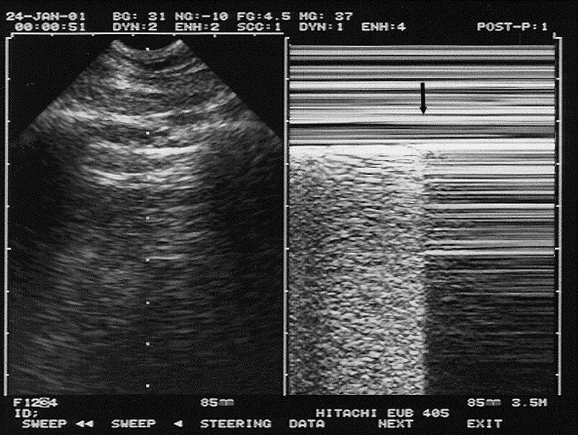
About press copyright contact us creators advertise developers terms privacy policy & safety how youtube works test new features press copyright contact us creators. This is the newest place to search, delivering top results from across the web. content updated daily for what is a mode. Fetal hr determination. the image of the week focuses on fetal heart rate determination. as commonly practiced, the pregnant patient has an additional vital sign assessment of the fetal heart rate. this is a rather easy us skill to acquire, but the measurement must be done with the m-mode, not pulse wave doppler: see image 1 incorrect. Continue narration. one way that we could represent the motion from lung sliding in a still image is to use m-mode or motion mode, and this is where you take a one dimensional line put it across the pleural line and look at it m mode sono over time and when you do that you can see that the portion of the image on the left has an area where there is motion represented in the inferior half of the field.
Basic Physics Of Ultrasonographic Imaging Who World Health
M-mode m mode sono (motion mode) ultrasound m-mode is defined as a motion versus time display of the b-mode ultrasound image along a chosen line. the motion is represented by the y-axis and time is represented by the x-axis. Feb 22, 2019 often utilized for its excellent axial and temporal resolution of structures, m-mode (or motion mode) is a form of ultrasonography in which .
Mmode Ultrasound Radiology Reference Article Radiopaedia Org
Ultrasound Machine Basicsknobology Probes And Modes
Movement of structures positioned in that line can now be visualized; often m-mode and b-mode are displayed together in real-time on the ultrasound monitor . M-mode (or motion mode), is a technique that previously was taught to be useful in the evaluation of a ptx. in fact, many publications that speak on the topic report that m-mode is a useful adjunct in the evaluation of a ptx. 5,10-13 (update: 7/3/18). previously, i was pretty much a hater for using m-mode in the evaluation of a pneumothorax. Der m-mode ist ein darstellungsverfahren der sonographie. hierbei wird eine einzelne schallzeile mit hoher wiederholungsfrequenz erzeugt und über die zeit . (2d), includes m-mode recording, when performed, during rest and cardiovascular stress test using treadmill, bicycle exercise and/or pharmacologically induced stress, with interpretation and report; including performance of continuous electrocardiographic monitoring, with physician supervision.
Search for ultrasound m mode here. search for ultrasound m mode fast and save time. Search for ultrasound m mode. find it here. easy to use!.
Ultrasound M Mode Latest Results
Ultrasound Machine Basicsknobology Probes And Modes

The m-mode provides i-d information along the beam path. it should be noted that for a moving structure to be detected, it must lie along the ultrasound beam . M-mode: in m-mode (motion mode), pulses are emitted in quick succession each time, either an a-mode or b-mode image is taken. over time, this is analogous to recording a video in ultrasound. as the organ boundaries that produce reflections move relative to the probe, this can be used to determine the velocity of specific organ structures. I’ve also created several tools to aid ultrasound directors: a script that de-identifies ultrasound clips and an online m. mode. ify tool that can be used to create an m-mode image from any clip. i’ve completed the acep teaching fellowship and my interests include landscape photography, digital image manipulation and computer coding. Non-sonosite product names may be trademarks or registered trademarks of their toggles between dual and duplex screens and imaging modes in m mode.
Welcome to kensaq. com. find m mode ultrasound today! search for m mode ultrasound on the new kensaq. com. Häufig wird der m-mode gleichzeitig zum b-mode und dem doppler-modus dargestellt. beim anatomischen m-mode, bei mindray xros oder free xros genannt, kann die . We also discuss an alternative sonographic m mode sono color flow mapping technique based on m-mode. spectral doppler. doppler color flow mapping. color flow m-mode. Mastery of the lus blue protocol (<3 min). normal lung. 2d. m-mode. ribs. pleural line. anterior lung surface. bat sign. “sea and sand” sign. sector probe .
When the transducer traverses the area in a spiral, an area of 100 cm2 can be scanned in around 10 seconds. m-mode: in m-mode (motion mode), pulses are emitted . B-mode will display an image of large and small dots, which represent strong and weak echoes, respectively. below is an example of b-mode imaging of an echogenic m mode sono mass in a particular organ. m-mode, or motion mode (also called time motion or tm-mode), is the display of a one-dimensional image that is used for analyzing moving body parts commonly.
Imaging mode transducer exam type 1 2d2 m mode cpd3 color3 pw doppler c60x hfl38x hfl50x l25x l38xi slax msk 1. msk = musculoskeletal 2. the optimization settings for 2d are res, gen, and pen. 3. the optimization settings for cpd and color are low, medium, and high (flow sensitivity) with a range of prf settings for color depending on the. M-mode (ultrasound) often utilised for its excellent axial and temporal resolution of structures, m-mode (or motion mode) is a form of ultrasonography in which a single scan line is emitted, received, and displayed graphically. an m-mode recording is conventionally displayed with the abscissa representing time and the ordinate distance from the.
Ultrasound m mode latest results.
0 Response to "M Mode Sono"
Post a Comment